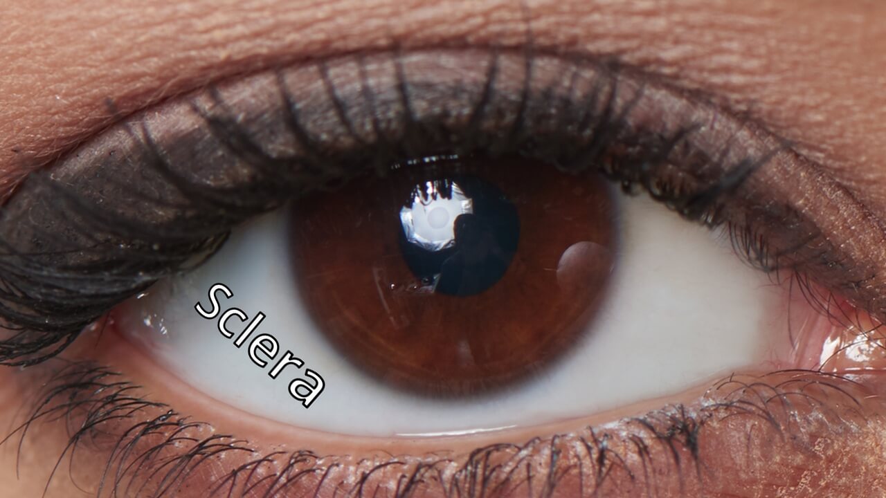
Sclera Is Derived From The Greek Word Meaning - All six ocular muscles insert into the sclera. The sclera is the white, outermost layer of the eyeball. To address this gap, we conducted a comprehensive investigation using ultrasonic elastography, optical coherence elastography, and. The in vivo biaxial stress state of the sclera. Sclera is an opaque, elastic, and resilient tissue of the eye. The sclera and the cornea form the rigid outer covering of the eye. During biaxial tensile testing, the spherical sclera is flattened and cut into a planar square specimen.
Sclera is an opaque, elastic, and resilient tissue of the eye. The in vivo biaxial stress state of the sclera. The sclera and the cornea form the rigid outer covering of the eye. To address this gap, we conducted a comprehensive investigation using ultrasonic elastography, optical coherence elastography, and. The sclera is the white, outermost layer of the eyeball. During biaxial tensile testing, the spherical sclera is flattened and cut into a planar square specimen. All six ocular muscles insert into the sclera.
All six ocular muscles insert into the sclera. To address this gap, we conducted a comprehensive investigation using ultrasonic elastography, optical coherence elastography, and. During biaxial tensile testing, the spherical sclera is flattened and cut into a planar square specimen. The sclera and the cornea form the rigid outer covering of the eye. Sclera is an opaque, elastic, and resilient tissue of the eye. The in vivo biaxial stress state of the sclera. The sclera is the white, outermost layer of the eyeball.

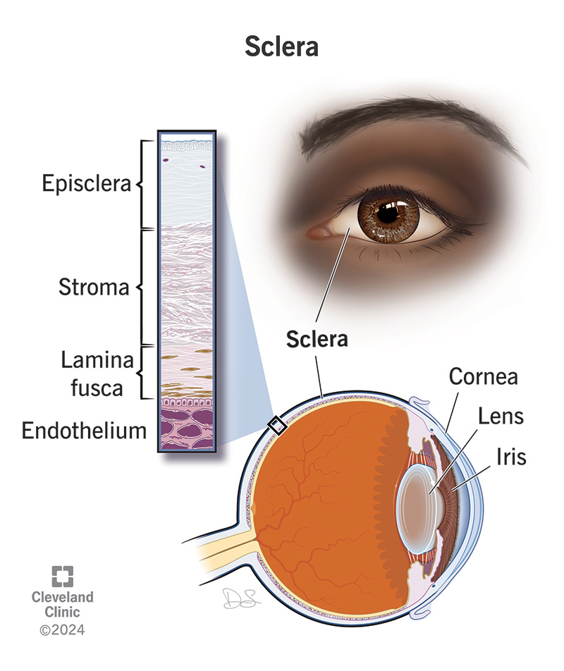
Episclera Scleritis And Episcleritis
The sclera and the cornea form the rigid outer covering of the eye. To address this gap, we conducted a comprehensive investigation using ultrasonic elastography, optical coherence elastography, and. Sclera is an opaque, elastic, and resilient tissue of the eye. During biaxial tensile testing, the spherical sclera is flattened and cut into a planar square specimen. The in vivo biaxial.
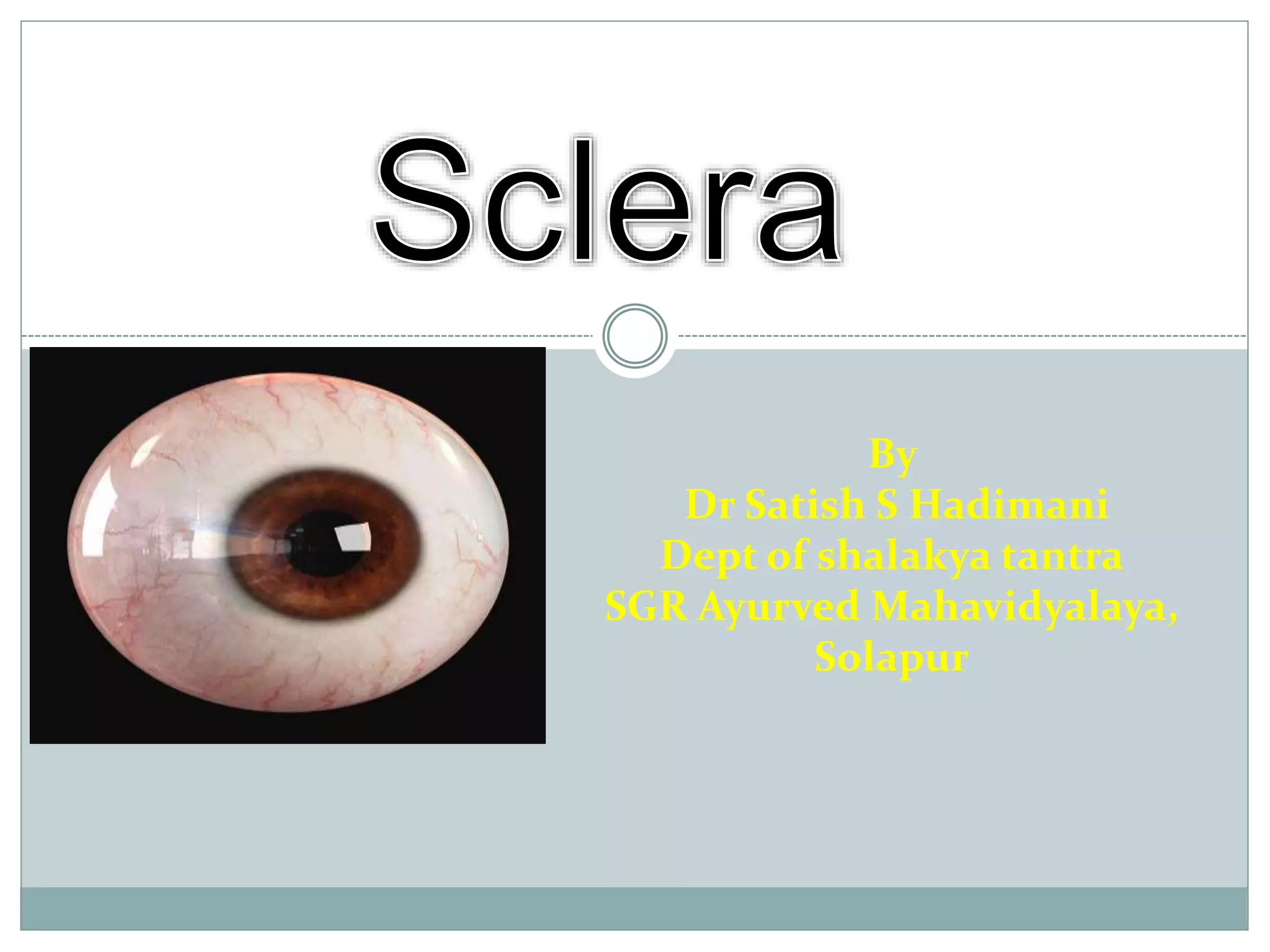
Anatomy of cornea and sclera PPTX Eye and Vision Conditions
The sclera and the cornea form the rigid outer covering of the eye. The sclera is the white, outermost layer of the eyeball. Sclera is an opaque, elastic, and resilient tissue of the eye. All six ocular muscles insert into the sclera. To address this gap, we conducted a comprehensive investigation using ultrasonic elastography, optical coherence elastography, and.

Ophthalmology, Endocrinology, and Medical Specialties ppt download
The sclera and the cornea form the rigid outer covering of the eye. All six ocular muscles insert into the sclera. During biaxial tensile testing, the spherical sclera is flattened and cut into a planar square specimen. Sclera is an opaque, elastic, and resilient tissue of the eye. To address this gap, we conducted a comprehensive investigation using ultrasonic elastography,.

Sclera (Human Anatomy) Image, Functions, Diseases and Treatments
The sclera and the cornea form the rigid outer covering of the eye. All six ocular muscles insert into the sclera. The sclera is the white, outermost layer of the eyeball. To address this gap, we conducted a comprehensive investigation using ultrasonic elastography, optical coherence elastography, and. The in vivo biaxial stress state of the sclera.

Unveiling The Secrets Of The White Underneath A Comprehensive Guide To
The sclera is the white, outermost layer of the eyeball. The in vivo biaxial stress state of the sclera. During biaxial tensile testing, the spherical sclera is flattened and cut into a planar square specimen. All six ocular muscles insert into the sclera. Sclera is an opaque, elastic, and resilient tissue of the eye.
Understanding The Sclera In Eye Anatomy Deliver Contacts
The sclera and the cornea form the rigid outer covering of the eye. Sclera is an opaque, elastic, and resilient tissue of the eye. To address this gap, we conducted a comprehensive investigation using ultrasonic elastography, optical coherence elastography, and. The sclera is the white, outermost layer of the eyeball. The in vivo biaxial stress state of the sclera.

Sclera Anatomy, Function, and Related Eye Problems
The sclera is the white, outermost layer of the eyeball. Sclera is an opaque, elastic, and resilient tissue of the eye. The in vivo biaxial stress state of the sclera. All six ocular muscles insert into the sclera. To address this gap, we conducted a comprehensive investigation using ultrasonic elastography, optical coherence elastography, and.

Sclera Anatomy, Function, and Related Eye Problems
To address this gap, we conducted a comprehensive investigation using ultrasonic elastography, optical coherence elastography, and. During biaxial tensile testing, the spherical sclera is flattened and cut into a planar square specimen. The sclera is the white, outermost layer of the eyeball. The sclera and the cornea form the rigid outer covering of the eye. Sclera is an opaque, elastic,.
Sclera Definition, Anatomy Function, 47 OFF
The sclera and the cornea form the rigid outer covering of the eye. The sclera is the white, outermost layer of the eyeball. All six ocular muscles insert into the sclera. To address this gap, we conducted a comprehensive investigation using ultrasonic elastography, optical coherence elastography, and. The in vivo biaxial stress state of the sclera.
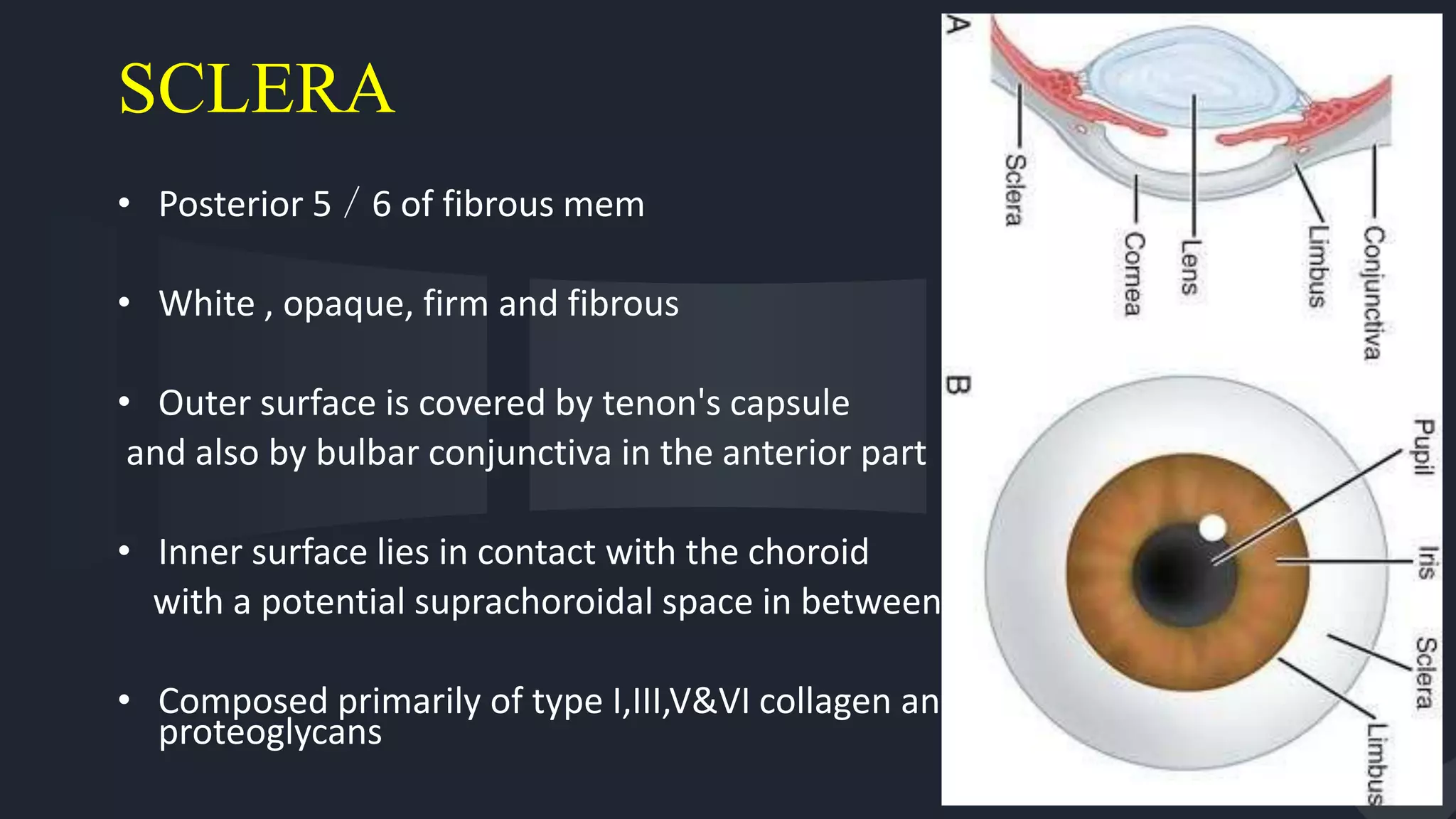
Anatomy of Sclera PPTX
The sclera is the white, outermost layer of the eyeball. The in vivo biaxial stress state of the sclera. Sclera is an opaque, elastic, and resilient tissue of the eye. To address this gap, we conducted a comprehensive investigation using ultrasonic elastography, optical coherence elastography, and. The sclera and the cornea form the rigid outer covering of the eye.
During Biaxial Tensile Testing, The Spherical Sclera Is Flattened And Cut Into A Planar Square Specimen.
To address this gap, we conducted a comprehensive investigation using ultrasonic elastography, optical coherence elastography, and. All six ocular muscles insert into the sclera. Sclera is an opaque, elastic, and resilient tissue of the eye. The sclera is the white, outermost layer of the eyeball.
The Sclera And The Cornea Form The Rigid Outer Covering Of The Eye.
The in vivo biaxial stress state of the sclera.